Taylor Swift made it again for 8 weeks #1 in the chart..Congrates Taylor..can't wait CSI and Teen Vogue (March)..

.jpg)
.jpg)
.jpg)
.jpg)
.jpg)


 result!


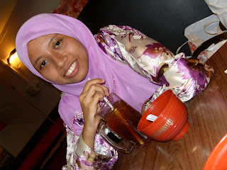
 yesterday, I went to Teppanyaki restaurant with Shiq & Elli. we were really surprise becoz the restaurant was really empty without any customers..hihi..the food was really nice in Teppanyaki but a bit spicy and I really can't handle spicy food a lot.. makes me dizzy and sweat..fuhh..we ordered ala carte menu which we enjoy Pari, Sotong and Lala set plus delicious mango jelly with cool ice lemon tea.. really big mug n I can't finished it.. =(..


